the eucharist
Throughout Church history, consecrated communion hosts have reportedly transformed into visible human flesh and blood. Modern laboratory analyses performed on these hosts across different continents have yielded consistent histological findings.
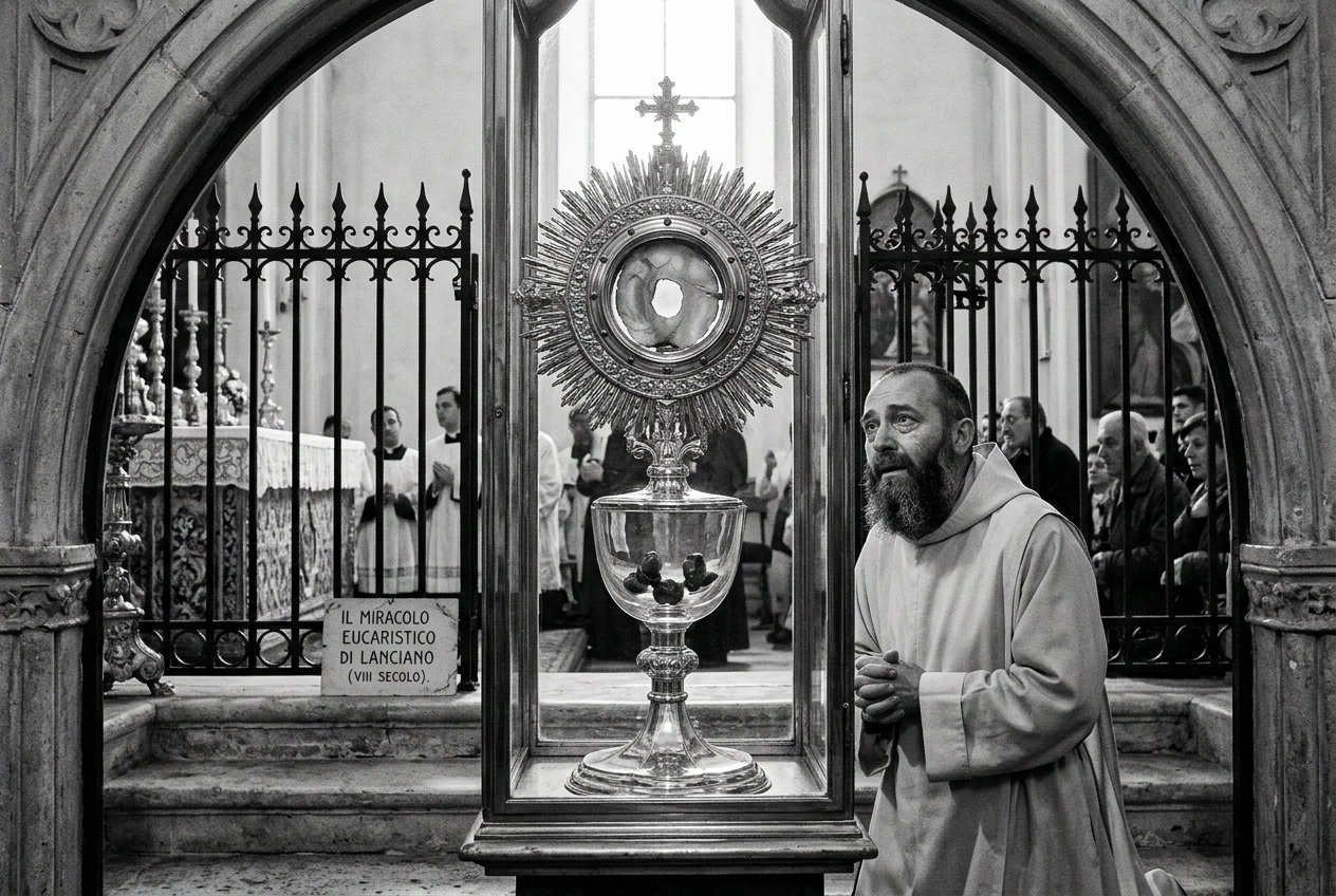
Histological Patterns Across Eucharistic Miracles
Tissue Type: Human myocardial (heart) tissue from the left ventricle wall.
White Blood Cells: Presence of active, living white blood cells (leukocytes) indicating severe physical trauma before tissue death.
Blood Typing: Historical tests consistently indicated Type AB blood.
Key Scientific Case Studies & Lab Results
Lanciano, Italy (8th Century Laboratory Findings)
In 1970–1971, Dr. Odoardo Linoli, Professor of Pathological Anatomy, conducted a scientific examination of the historical 8th-century Eucharistic miracle at Lanciano. His published analysis confirmed that the tissue consists of real human heart muscle (myocardium) and human blood.
Note on Scientific Accuracy: Prior claims regarding a secondary WHO/UN investigation have been debunked as an unverified rumor and are retracted from modern evidentiary claims.
Buenos Aires, Argentina (1996 Forensic Pathology)
In 1996, a discarded host found in a Buenos Aires parish transformed into a bloody substance. Cardinal Jorge Mario Bergoglio (later Pope Francis) commissioned a scientific study.
Samples sent blind to forensic pathologists (including Dr. Frederic Zugibe of Columbia University) revealed human heart tissue from the left ventricle wall and infiltrated white blood cells, indicating the individual was alive when the tissue sample was taken and had suffered severe chest trauma.
Sokółka & Legnica, Poland (2008–2013 Studies)
Independent histological studies conducted by the University of Białystok and the Department of Forensic Medicine in Wrocław confirmed that altered hosts in Sokółka and Legnica contained human cardiac tissue intertwined with bread fibers at a structural, non-artificial level.